 |
||||||||||||||||||||||||||||
 |
||||||||||||||||||||||||||||
| DP00062: Retinoic acid receptor RXR-alpha |  |
| General information | |
| DisProt: | DP00062 |
| Name: | Retinoic acid receptor RXR-alpha |
| Synonym(s): | RXRA_HUMAN Retinoid X receptor alpha Nuclear receptor subfamily 2 group B member 1 |
| First appeared in release: | Release 1.0 (08/01/2003) |
| UniProt: | P19793 |
| UniGene: | Hs.590886 |
| SwissProt: | RXRA_HUMAN |
| TrEMBL: | |
| NCBI (GI): | 133701 |
| Source organism: | Homo sapiens (Human) |
| Sequence length: | 462 |
| Percent disordered: | 6% |
| Homologues: | |
| Native sequence |
|
| Functional narrative |
Retinoid X receptor (RXR) is a member of the nuclear hormone receptor superfamily of transcription regulators. RXR plays a central role in the retinoid and non-steroid signaling pathways. RXR consists of three domains: an N-terminal region of unknown structure, a DNA binding domain (DBD) and a ligand-binding region. Binding occurs cooperatively as dimers to hormone response elements composed of two copies of the consensus half-site sequences of direct repeats of AGGTCA. When RXR binds 9-cis-retinoic acid it transactivates as a homodimer. The flexibility in the dimerization region allows different protein-protein interactions and response elements to be recognized. The order-disorder transition of the C-terminal DNA-binding domain extends the backbone to allow optimal interactions with the upstream DBD in the dimer. RXR is highly expressed in the liver and also found in the lung, kidney and heart. |
| Map of ordered and disordered regions | |
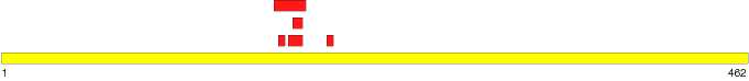  Note: 'Mouse' over a region to see the start and stop residues. Click on a region to see detailed information. | |
| Region 1 | |
| Type: | Disordered - Extended |
| Name: | D-BOX |
| Location: | 172 - 176 |
| Length: | 5 |
| Region sequence: | RDNKD |
| Modification type: | Engineered |
| PDB: | 1RXR:1 |
| Structural/functional type: | |
| Functional classes: | Molecular assembly |
| Functional subclasses: | Protein-protein binding Protein-DNA binding |
Detection methods:
| |
References:
| |
| Comments: | |
| Region 2 | |
| Type: | Disordered - Extended |
| Name: | Second Zinc Finger Binding Domain |
| Location: | 178 - 187 |
| Length: | 10 |
| Region sequence: | LIDKRQRNRC |
| Modification type: | Engineered |
| PDB: | 1RXR:1 |
| Structural/functional type: | Function arises via a disorder to order transition |
| Functional classes: | Molecular assembly |
| Functional subclasses: | Metal binding Protein-DNA binding |
Detection methods:
| |
References:
| |
| Comments: | |
| Region 3 | |
| Type: | Disordered - Extended |
| Name: | |
| Location: | 169 - 189 |
| Length: | 21 |
| Region sequence: | YTCRDNKDCLIDKRQRNRCQY |
| Modification type: | Engineered |
| PDB: | IRXR:1 |
| Structural/functional type: | Function arises via a disorder to order transition |
| Functional classes: | Molecular assembly |
| Functional subclasses: | Metal binding Protein-DNA binding |
Detection methods:
| |
References:
| |
Comments:This region contains the D-box as well as the second zinc binding domain and lies between the first and second helices. | |
| Region 4 | |
| Type: | Disordered - Extended |
| Name: | Second Zinc binding domain |
| Location: | 181 - 187 |
| Length: | 7 |
| Region sequence: | KRQRNRC |
| Modification type: | Engineered |
| PDB: | 1RXR:1 |
| Structural/functional type: | Function arises via a disorder to order transition |
| Functional classes: | Molecular assembly |
| Functional subclasses: | Metal binding Protein-DNA binding |
Detection methods:
| |
References:
| |
| Comments: | |
| Region 5 | |
| Type: | Disordered - Extended |
| Name: | C-terminal helix |
| Location: | 202 - 206 |
| Length: | 5 |
| Region sequence: | REAVQ |
| Modification type: | Engineered |
| PDB: | 1RXR:1 |
| Structural/functional type: | Function arises via an order to disorder transition |
| Functional classes: | Molecular assembly |
| Functional subclasses: | Protein-DNA binding Substrate/ligand binding Protein-protein binding |
Detection methods:
| |
References:
| |
Comments:The C-terminal helix undergoes an order to disorder transition upon binding to DNA. The unwinding of the helix most likely facilitates homodimer formation by maximizing interactions between the two DNA-bound RXR proteins. | |
| References | |
|
| If you have any comments or wish to provide additional references to this protein or its disordered region(s), please click here to e-mail us. |
 |
 |
 |
 |
 |
 |
|
|
|