| General information | | DisProt: | DP00207 | | Name: | Ribonuclease E | | Synonym(s): | RNE_ECOLI
RNase E
| | First appeared in release: | Release 2.1 (03/14/2005) | | UniProt: | P21513 | | UniGene: | | | SwissProt: | RNE_ECOLI | | TrEMBL: | | | NCBI (GI): | 34395929 | | Source organism: | Escherichia coli | | Sequence length: | 1061 | | Percent disordered: | 53% | | Homologues: | |
| Native sequence |
10 20 30 40 50 60
| | | | | |
MKRMLINATQ QEELRVALVD GQRLYDLDIE SPGHEQKKAN IYKGKITRIE PSLEAAFVDY - 60
GAERHGFLPL KEIAREYFPA NYSAHGRPNI KDVLREGQEV IVQIDKEERG NKGAALTTFI - 120
SLAGSYLVLM PNNPRAGGIS RRIEGDDRTE LKEALASLEL PEGMGLIVRT AGVGKSAEAL - 180
QWDLSFRLKH WEAIKKAAES RPAPFLIHQE SNVIVRAFRD YLRQDIGEIL IDNPKVLELA - 240
RQHIAALGRP DFSSKIKLYT GEIPLFSHYQ IESQIESAFQ REVRLPSGGS IVIDSTEALT - 300
AIDINSARAT RGGDIEETAF NTNLEAADEI ARQLRLRDLG GLIVIDFIDM TPVRHQRAVE - 360
NRLREAVRQD RARIQISHIS RFGLLEMSRQ RLSPSLGESS HHVCPRCSGT GTVRDNESLS - 420
LSILRLIEEE ALKENTQEVH AIVPVPIASY LLNEKRSAVN AIETRQDGVR CVIVPNDQME - 480
TPHYHVLRVR KGEETPTLSY MLPKLHEEAM ALPSEEEFAE RKRPEQPALA TFAMPDVPPA - 540
PTPAEPAAPV VAPAPKAAPA TPAAPAQPGL LSRFFGALKA LFSGGEETKP TEQPAPKAEA - 600
KPERQQDRRK PRQNNRRDRN ERRDTRSERT EGSDNREENR RNRRQAQQQT AETRESRQQA - 660
EVTEKARTAD EQQAPRRERS RRRNDDKRQA QQEAKALNVE EQSVQETEQE ERVRPVQPRR - 720
KQRQLNQKVR YEQSVAEEAV VAPVVEETVA AEPIVQEAPA PRTELVKVPL PVVAQTAPEQ - 780
QEENNADNRD NGGMPRRSRR SPRHLRVSGQ RRRRYRDERY PTQSPMPLTV ACASPELASG - 840
KVWIRYPIVR PQDVQVEEQR EQEEVHVQPM VTEVPVAAAI EPVVSAPVVE EVAGVVEAPV - 900
QVAEPQPEVV ETTHPEVIAA AVTEQPQVIT ESDVAVAQEV AEQAEPVVEP QEETADIEEV - 960
VETAEVVVAE PEVVAQPAAP VVAEVAAEVE TVAAVEPEVT VEHNHATAPM TRAPAPEYVP - 1020
EAPRHSDWQR PTFAFEGKGA AGGHTATHHA SAAPARPQPV E
|
| Functional narrative |
Ribonuclease E is made up of two domains, the N-terminal domain, NTD and the C-terminal domain, CTD. The CTD, which is disordered, functionally organizes the RNA degradosome (Callaghan 2004). Within the CTD, there are sites for self-association and interaction with helicase-RhlB, PNPase and RNA.
|
| Map of ordered and disordered regions |
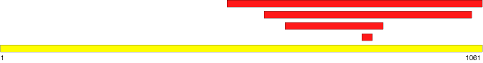

Note: 'Mouse' over a region to see the start and stop residues. Click on a region to see detailed information.
|
| Region 1 | | Type: | Disordered | | Name: | CTD (C-terminal domain) | | Location: | 499 - 1061 | | Length: | 563 | | Region sequence: |
SYMLPKLHEEAMALPSEEEFAERKRPEQPALATFAMPDVPPAPTPAEPAAPVVAPAPKAA
PATPAAPAQPGLLSRFFGALKALFSGGEETKPTEQPAPKAEAKPERQQDRRKPRQNNRRD
RNERRDTRSERTEGSDNREENRRNRRQAQQQTAETRESRQQAEVTEKARTADEQQAPRRE
RSRRRNDDKRQAQQEAKALNVEEQSVQETEQEERVRPVQPRRKQRQLNQKVRYEQSVAEE
AVVAPVVEETVAAEPIVQEAPAPRTELVKVPLPVVAQTAPEQQEENNADNRDNGGMPRRS
RRSPRHLRVSGQRRRRYRDERYPTQSPMPLTVACASPELASGKVWIRYPIVRPQDVQVEE
QREQEEVHVQPMVTEVPVAAAIEPVVSAPVVEEVAGVVEAPVQVAEPQPEVVETTHPEVI
AAAVTEQPQVITESDVAVAQEVAEQAEPVVEPQEETADIEEVVETAEVVVAEPEVVAQPA
APVVAEVAAEVETVAAVEPEVTVEHNHATAPMTRAPAPEYVPEAPRHSDWQRPTFAFEGK
GAAGGHTATHHASAAPARPQPVE | | Modification type: | Fragment
Native
| | PDB: | | | Structural/functional type: | Function arises from the disordered state
Function arises via a disorder to order transition
| | Functional classes: | Unknown
| | Functional subclasses: | Protein-rRNA binding
Autoregulatory
Protein-protein binding
| Detection methods:
- Sensitivity to proteolysis
- Circular dichroism (CD) spectroscopy, far-UV (275 K; collected between 185 nm and 260 nm (1 nm steps ))
- Circular dichroism (CD) spectroscopy, far-UV (295 K; collected between 185 nm and 260 nm (1 nm steps ))
- Circular dichroism (CD) spectroscopy, far-UV (315 K; collected between 185 nm and 260 nm (1 nm steps ))
- Circular dichroism (CD) spectroscopy, far-UV (335 K; collected between 185 nm and 260 nm (1 nm steps ))
- Circular dichroism (CD) spectroscopy, far-UV (355 K; collected between 185 nm and 260 nm (1 nm steps ))
- Small-angle X-ray scattering (SAXS)
| References:
- Callaghan AJ, Aurikko JP, Ilag LL, Gunter Grossmann J, Chandran V, Kuhnel K, Poljak L, Carpousis AJ, Robinson CV, Symmons MF, Luisi BF. "Studies of the RNA degradosome-organizing domain of the Escherichia coli ribonuclease RNase E." J Mol Biol. 2004; 340(5): 965-79. PubMed: 15236960
| Comments:CTD can be cross-linked to itself, thus forming dimers. The region that can self-interact is from 500-752 residues.
Within the disordered CTD, there are four regions that have predicted structural propensity. These segments are called RISPs (regions of increased structural propensity). The RISPs are; segment A- residues 565-585, segment B- residues 633-712, segment C- residues 839-850 and segment D- residues 1021-1061.
|
| Region 2 | | Type: | Disordered | | Name: | R-domain | | Location: | 628 - 843 | | Length: | 216 | | Region sequence: |
ERTEGSDNREENRRNRRQAQQQTAETRESRQQAEVTEKARTADEQQAPRRERSRRRNDDK
RQAQQEAKALNVEEQSVQETEQEERVRPVQPRRKQRQLNQKVRYEQSVAEEAVVAPVVEE
TVAAEPIVQEAPAPRTELVKVPLPVVAQTAPEQQEENNADNRDNGGMPRRSRRSPRHLRV
SGQRRRRYRDERYPTQSPMPLTVACASPELASGKVW | | Modification type: | Engineered
Fragment
| | PDB: | | | Structural/functional type: | Function arises from the disordered state
| | Functional classes: | Unknown
| | Functional subclasses: | Protein-rRNA binding
Protein-protein binding
| Detection methods:
- Circular dichroism (CD) spectroscopy, far-UV
- Size exclusion/gel filtration chromatography
| References:
- Callaghan AJ, Aurikko JP, Ilag LL, Gunter Grossmann J, Chandran V, Kuhnel K, Poljak L, Carpousis AJ, Robinson CV, Symmons MF, Luisi BF. "Studies of the RNA degradosome-organizing domain of the Escherichia coli ribonuclease RNase E." J Mol Biol. 2004; 340(5): 965-79. PubMed: 15236960
| Comments:The experimental domain included the coiled coil segment B region.
The 7SrRNA binding region is within this domain.
There is an arginine-rich region within this domain, residues 796-819, that may gain some structure upon the binding to RNA.
This domain contains the portion of the disordered CTD that binds helicase RhlB. It binds helicase RhlB in a 1:1 ratio. The binding of helicase RhlB to this domain stimulates the ATPase activity of the helicase. Binding to the helicase protein does not induce folding of the R-domain.
|
| Region 3 | | Type: | Disordered - Extended | | Name: | | | Location: | 796 - 819 | | Length: | 24 | | Region sequence: |
RRSRRSPRHLRVSGQRRRRYRDER | | Modification type: | Fragment
| | PDB: | | | Structural/functional type: | Function arises via a disorder to order transition
| | Functional classes: | Unknown
| | Functional subclasses: | Protein-rRNA binding
| Detection methods:
| References:
- Callaghan AJ, Aurikko JP, Ilag LL, Gunter Grossmann J, Chandran V, Kuhnel K, Poljak L, Carpousis AJ, Robinson CV, Symmons MF, Luisi BF. "Studies of the RNA degradosome-organizing domain of the Escherichia coli ribonuclease RNase E." J Mol Biol. 2004; 340(5): 965-79. PubMed: 15236960
| Comments:This region is completely contained within the R-domain.
This region is disordered because it is within the CTD domain of the protein but it is thought to gain structure upon the binding of RNA.
|
| Region 4 | | Type: | Disordered - Extended | | Name: | native-CTD | | Location: | 580 - 1038 | | Length: | 459 | | Region sequence: |
ALFSGGEETKPTEQPAPKAEAKPERQQDRRKPRQNNRRDRNERRDTRSERTEGSDNREEN
RRNRRQAQQQTAETRESRQQAEVTEKARTADEQQAPRRERSRRRNDDKRQAQQEAKALNV
EEQSVQETEQEERVRPVQPRRKQRQLNQKVRYEQSVAEEAVVAPVVEETVAAEPIVQEAP
APRTELVKVPLPVVAQTAPEQQEENNADNRDNGGMPRRSRRSPRHLRVSGQRRRRYRDER
YPTQSPMPLTVACASPELASGKVWIRYPIVRPQDVQVEEQREQEEVHVQPMVTEVPVAAA
IEPVVSAPVVEEVAGVVEAPVQVAEPQPEVVETTHPEVIAAAVTEQPQVITESDVAVAQE
VAEQAEPVVEPQEETADIEEVVETAEVVVAEPEVVAQPAAPVVAEVAAEVETVAAVEPEV
TVEHNHATAPMTRAPAPEYVPEAPRHSDWQRPTFAFEGK | | Modification type: | Complex
Fragment
| | PDB: | | | Structural/functional type: | Function arises from the disordered state
| | Functional classes: | Unknown
| | Functional subclasses: | Protein-protein binding
| Detection methods:
- Sensitivity to proteolysis (Complex, (native-CTD-enolase) less sensitive to chymotrypsin; Complex, (native-CTD-enolase) very sensitive to Protease K)
| References:
- Callaghan AJ, Aurikko JP, Ilag LL, Gunter Grossmann J, Chandran V, Kuhnel K, Poljak L, Carpousis AJ, Robinson CV, Symmons MF, Luisi BF. "Studies of the RNA degradosome-organizing domain of the Escherichia coli ribonuclease RNase E." J Mol Biol. 2004; 340(5): 965-79. PubMed: 15236960
| Comments:This domain contains the entire C segment, residues 839-850.
This fragment is a version of CTD isolated under native conditions without affinity tags.
This domain was co-purified with enolase. The binding region included residues 816-1038.
|
| References |
- Callaghan AJ, Aurikko JP, Ilag LL, Gunter Grossmann J, Chandran V, Kuhnel K, Poljak L, Carpousis AJ, Robinson CV, Symmons MF, Luisi BF. "Studies of the RNA degradosome-organizing domain of the Escherichia coli ribonuclease RNase E." J Mol Biol. 2004; 340(5): 965-79. PubMed: 15236960
- Kaberdin VR, Miczak A, Jakobsen JS, Lin-Chao S, McDowall KJ, von Gabain A. "The endoribonucleolytic N-terminal half of Escherichia coli RNase E is evolutionarily conserved in Synechocystis sp. and other bacteria but not the C-terminal half, which is sufficient for degradosome assembly." Proc Natl Acad Sci U S A. 1998; 95(20): 11637-11642. PubMed: 9751718
- Leroy A, Vanzo NF, Sousa S, Dreyfus M, Carpousis AJ. "Function in Escherichia coli of the non-catalytic part of RNase E: role in the degradation of ribosome-free mRNA." Mol Microbiol. 2002; 45(5): 1231-1243. PubMed: 12207692
- Vanzo NF, Li YS, Py B, Blum E, Higgins CF, Raynal LC, Krisch HM, Carpousis AJ. "Ribonuclease E organizes the protein interactions in the Escherichia coli RNA degradosome." Genes Dev. 1998; 12(17): 2770-2781. PubMed: 9732274
|
| Comments |
PDB entries 1SLJ:A, 1SMX:A, B, and 1SN8:A, B pertain to the ordered region of RNase E. These entries are related to amino acid residues 35-125.
|
| If you have any comments or wish to provide additional references to this protein or its disordered region(s), please click here to e-mail us. |
Disprot-footer
|


