| General information | | DisProt: | DP00680 | | Name: | Trypsin inhibitor 2 | | Synonym(s): | ITR2_MOMCO
Macrocyclic trypsin inhibitor II
MCoTI-II
Trypsin inhibitor II
| | First appeared in release: | Release 5.1 (05/28/2010) | | UniProt: | P82409 | | UniGene: | | | SwissProt: | ITR2_MOMCO | | TrEMBL: | | | NCBI (GI): | 547744 | | Source organism: | Momordica cochinchinensis (Spiny bitter cucumber) | | Sequence length: | 34 | | Percent disordered: | 44% | | Homologues: | |
| Native sequence |
10 20 30 40 50 60
| | | | | |
SGSDGGVCPK ILKKCRRDSD CPGACICRGN GYCG
|
| Functional narrative |
Inhibits trypsin; probably participates in a plant defense mechanism.
Secreted. The presence of a 'disulfide through disulfide knot' structurally defines this protein as a knottin. A cyclic succinimide probably forms by loss of water between Asp-4 and Gly-5, that can then rehydrate to either the original peptide bond or to a beta-aspartyl isopeptide bond. Three isoforms of MCoTI-II are detected, two with the parent molecular weight, corresponding to the unmodified and proposed isopeptide forms, and one with a molecular weight 18 Da lower, corresponding to a succinimide cross-linked form.
This is a cyclic peptide. Belongs to the protease inhibitor I7 (squash-type serine protease inhibitor) family. (UniProt)
----------------------------------------
Protein exists as a circular protein. Residue G34 links to residue S1. Amino acid sequence of this protein is numbered in such a way as to match sequence of its linear homologues.
----------------------------------------
MCoTI-II shares structural similarity as well as high sequence identity with other linear squash trypsin inhibitors, but has low sequence identity with other cyclic knottins, in which only the cysteines (and consequently the disulfide bridges) are conserved. Cyclization likely contributes to the inhibition of degradation by exoproteases. (Felizmenio-Quimio et al , 2001; Heitz et al, 2001)
|
| Map of ordered and disordered regions |
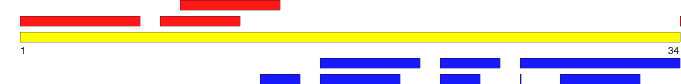

Note: 'Mouse' over a region to see the start and stop residues. Click on a region to see detailed information.
|
| Region 1 | | Type: | Disordered | | Name: | C-to-N linker, Loop 6 [cyclizing residue link to Rgn2] | | Location: | 34 - 34 | | Length: | 1 | | Region sequence: |
G | | Modification type: | Native
| | PDB: | 1HA9:A, 1IB9:A | | Structural/functional type: | Function arises from the disordered state
| | Functional classes: | | | Functional subclasses: | Intraprotein interaction
Flexible linkers/spacers
| Detection methods:
- Nuclear magnetic resonance (NMR) (288 K; pH: 3.5; MCoTI-II 1 mM; 90% H2O, 10% D2O (v/v. Or 99.99% D2O); also performed at 293K and 298K)
- Nuclear magnetic resonance (NMR) (285 K; pH: 3.4; 90% H2O, 10% D2O (v/v. Or 100% D2O); also performed at 300K; MCoTI-II 2.5 mM)
| References:
- Felizmenio-Quimio ME, Daly NL, Craik DJ. "Circular proteins in plants: solution structure of a novel macrocyclic trypsin inhibitor from Momordica cochinchinensis." J Biol Chem. 2001; 276(25): 22875-82. PubMed: 11292835
- Heitz A, Hernandez JF, Gagnon J, Hong TT, Pham TT, Nguyen TM, Le-Nguyen D, Chiche L. "Solution structure of the squash trypsin inhibitor MCoTI-II. A new family for cyclic knottins." Biochemistry. 2001; 40(27): 7973-83. PubMed: 11434766
| Comments:Protein exists as a circular protein. Residue G34 links to residue S1. Amino acid sequence of this protein is numbered in such a way as to match sequence of its highly-conserved linear homologues.
----------
PDB structure 1HA9 is from Heitz et al (2001), PDB structure 1IB9 is from Felizmenio-Quimio et al (2001). See overall protein comments for putative cysteine bonding pattern that forms knotted structure.
----------
Regarding Regions 1 and 2, Heitz et al (2001) call this circularizing segment consisting of aa G34 to V7 “C-to-N linker,” while Felizmenio-Quimio et al (2001) refer to it as “Loop 6.”
----------
Regions 3 through 7 are from Heitz et al (2001); Regions 8 through 12 are from Felizmenio-Quimio et al (2001) and exclude the cysteines which bond to form the knottin-type structure..
Loop 6 (Felizmenio-Quimio et al (2001)) is preceded by C33 (Cys VI), which bonds with C21 (Cys III).
|
| Region 2 | | Type: | Disordered | | Name: | C-to-N linker, Loop 6 [cyclized w/Rgn 1] | | Location: | 1 - 7 | | Length: | 7 | | Region sequence: |
SGSDGGV | | Modification type: | Native
| | PDB: | 1HA9:A, 1IB9:A | | Structural/functional type: | Function arises from the disordered state
| | Functional classes: | | | Functional subclasses: | Intraprotein interaction
Flexible linkers/spacers
| Detection methods:
- Nuclear magnetic resonance (NMR) (288 K; pH: 3.5; 90% H2O, 10% D2O (v/v. Or 99.99% D2O); also performed at 293K and 298K; MCoTI-II 1 mM)
- Nuclear magnetic resonance (NMR) (285 K; pH: 3.4; 90% H2O, 10% D2O (v/v. Or 100% D2O); also performed at 300K; MCoTI-II)
| References:
- Felizmenio-Quimio ME, Daly NL, Craik DJ. "Circular proteins in plants: solution structure of a novel macrocyclic trypsin inhibitor from Momordica cochinchinensis." J Biol Chem. 2001; 276(25): 22875-82. PubMed: 11292835
- Heitz A, Hernandez JF, Gagnon J, Hong TT, Pham TT, Nguyen TM, Le-Nguyen D, Chiche L. "Solution structure of the squash trypsin inhibitor MCoTI-II. A new family for cyclic knottins." Biochemistry. 2001; 40(27): 7973-83. PubMed: 11434766
| Comments:Protein exists as a circular protein. Residue G34 links to residue S1. Amino acid sequence of this protein is numbered in such a way as to match sequence of its highly-conserved linear homologues.
----------
PDB structure 1HA9 is from Heitz et al (2001), PDB structure 1IB9 is from Felizmenio-Quimio et al (2001). See overall protein comments for putative cysteine bonding pattern that forms knotted structure.
----------
Regarding Regions 1 and 2, Heitz et al (2001) call this circularizing segment consisting of aa G34 to V7 “C-to-N linker,” while Felizmenio-Quimio et al (2001) refer to it as “Loop 6.”
----------
Regions 3 through 7 are from Heitz et al (2001); Regions 8 through 12 are from Felizmenio-Quimio et al (2001) and exclude the cysteines which bond to form the knottin-type structure..
A contact is formed between G6 and A24, according to Felizmenio-Quimio et al (2001).
|
| Region 3 | | Type: | Disordered | | Name: | Inhibitory Loop | | Location: | 8 - 12 | | Length: | 5 | | Region sequence: |
CPKIL | | Modification type: | Native
| | PDB: | 1HA9:A | | Structural/functional type: | Function arises from the disordered state
| | Functional classes: | Molecular recognition effectors
| | Functional subclasses: | Regulation of proteolysis in vivo
| Detection methods:
- Nuclear magnetic resonance (NMR) (285 K; pH: 3.4; 90% H2O, 10% D2O (v/v. Or 100% D2O); also performed at 300K; MCoTI-II 2.5 mM)
| References:
- Heitz A, Hernandez JF, Gagnon J, Hong TT, Pham TT, Nguyen TM, Le-Nguyen D, Chiche L. "Solution structure of the squash trypsin inhibitor MCoTI-II. A new family for cyclic knottins." Biochemistry. 2001; 40(27): 7973-83. PubMed: 11434766
| Comments:
|
| Region 4 | | Type: | Ordered | | Name: | β-sheet | | Location: | 13 - 15 | | Length: | 3 | | Region sequence: |
KKC | | Modification type: | Native
| | PDB: | 1HA9:A | | Structural/functional type: | Relationship to function unknown
| | Functional classes: | Unknown
| | Functional subclasses: | Unknown
| Detection methods:
- Nuclear magnetic resonance (NMR) (285 K; pH: 3.4; 90% H2O, 10% D2O (v/v. Or 100% D2O); also performed at 300K; MCoTI-II 2.5 mM)
| References:
- Heitz A, Hernandez JF, Gagnon J, Hong TT, Pham TT, Nguyen TM, Le-Nguyen D, Chiche L. "Solution structure of the squash trypsin inhibitor MCoTI-II. A new family for cyclic knottins." Biochemistry. 2001; 40(27): 7973-83. PubMed: 11434766
| Comments:Heitz et al (2001) describe the presence of a motif common in squash trypsin inhibitors and cysteine-rich proteins. This motif is an anti-parallel triple-stranded β-sheet involving K13-C15, I26-R28 and Y32-G34.
|
| Region 5 | | Type: | Ordered | | Name: | 3-10 helix | | Location: | 16 - 21 | | Length: | 6 | | Region sequence: |
RRDSDC | | Modification type: | Native
| | PDB: | 1HA9:A | | Structural/functional type: | Relationship to function unknown
| | Functional classes: | Unknown
| | Functional subclasses: | Unknown
| Detection methods:
- Nuclear magnetic resonance (NMR) (285 K; pH: 3.4; 90% H2O, 10% D2O (v/v. Or 100% D2O); also performed at 300K; MCoTI-II 2.5 mM)
| References:
- Heitz A, Hernandez JF, Gagnon J, Hong TT, Pham TT, Nguyen TM, Le-Nguyen D, Chiche L. "Solution structure of the squash trypsin inhibitor MCoTI-II. A new family for cyclic knottins." Biochemistry. 2001; 40(27): 7973-83. PubMed: 11434766
| Comments:
|
| Region 6 | | Type: | Ordered | | Name: | β-turn | | Location: | 22 - 25 | | Length: | 4 | | Region sequence: |
PGAC | | Modification type: | Native
| | PDB: | 1HA9:A | | Structural/functional type: | Relationship to function unknown
| | Functional classes: | Unknown
| | Functional subclasses: | Unknown
| Detection methods:
- Nuclear magnetic resonance (NMR) (285 K; pH: 3.4; 90% H2O, 10% D2O (v/v. Or 100% D2O); also performed at 300K; MCoTI-II 2.5 mM)
| References:
- Heitz A, Hernandez JF, Gagnon J, Hong TT, Pham TT, Nguyen TM, Le-Nguyen D, Chiche L. "Solution structure of the squash trypsin inhibitor MCoTI-II. A new family for cyclic knottins." Biochemistry. 2001; 40(27): 7973-83. PubMed: 11434766
| Comments:
|
| Region 7 | | Type: | Ordered | | Name: | β-haripin | | Location: | 26 - 34 | | Length: | 9 | | Region sequence: |
ICRGNGYCG | | Modification type: | Native
| | PDB: | 1HA9:A | | Structural/functional type: | Relationship to function unknown
| | Functional classes: | Unknown
| | Functional subclasses: | Unknown
| Detection methods:
- Nuclear magnetic resonance (NMR) (285 K; pH: 3.4; 90% H2O, 10% D2O (v/v. Or 100% D2O); also performed at 300K; MCoTI-II 2.5 mM)
| References:
- Heitz A, Hernandez JF, Gagnon J, Hong TT, Pham TT, Nguyen TM, Le-Nguyen D, Chiche L. "Solution structure of the squash trypsin inhibitor MCoTI-II. A new family for cyclic knottins." Biochemistry. 2001; 40(27): 7973-83. PubMed: 11434766
| Comments:According to Heitz et al (2001), this β-hairpin segment is comprised of a β-sheet at I26-C27, a β-turn at R28-G31 and another β-sheet at Y32-G34. The protein is cyclical as G34 connects to S1.
Heitz et al (2001) describe the presence of a motif common in squash trypsin inhibitors and cysteine-rich proteins. This motif is an anti-parallel triple-stranded β-sheet involving K13-C15, I26-R28 and Y32-G34.
|
| Region 8 | | Type: | Disordered | | Name: | Loop 1 | | Location: | 9 - 14 | | Length: | 6 | | Region sequence: |
PKILKK | | Modification type: | Native
| | PDB: | 1IB9:A | | Structural/functional type: | Function arises from the disordered state
| | Functional classes: | Molecular recognition effectors
| | Functional subclasses: | Regulation of proteolysis in vivo
| Detection methods:
- Nuclear magnetic resonance (NMR) (288 K; pH: 3.5; 90% H2O, 10% D2O (v/v. Or 99.99% D2O); also performed at 293K and 298K; MCoTI-II 1 mM)
| References:
- Felizmenio-Quimio ME, Daly NL, Craik DJ. "Circular proteins in plants: solution structure of a novel macrocyclic trypsin inhibitor from Momordica cochinchinensis." J Biol Chem. 2001; 276(25): 22875-82. PubMed: 11292835
| Comments:Felizmenio-Quimio et al (2001) describe Loop 1 as having a 3+ charge, and as containing a putative active site at I11-L12. Loop 1 is preceded by C8, also know as Cys I (this author denotes cysteines with Roman numerals), which bonds with C25 (Cys IV).
|
| Region 9 | | Type: | Ordered | | Name: | Loop 2 | | Location: | 16 - 20 | | Length: | 5 | | Region sequence: |
RRDSD | | Modification type: | Native
| | PDB: | 1IB9:A | | Structural/functional type: | Relationship to function unknown
| | Functional classes: | Unknown
| | Functional subclasses: | Unknown
| Detection methods:
- Nuclear magnetic resonance (NMR) (288 K; pH: 3.5; 90% H2O, 10% D2O (v/v. Or 99.99% D2O); also performed at 293K and 298K; MCoTI-II 1 mM)
| References:
- Felizmenio-Quimio ME, Daly NL, Craik DJ. "Circular proteins in plants: solution structure of a novel macrocyclic trypsin inhibitor from Momordica cochinchinensis." J Biol Chem. 2001; 276(25): 22875-82. PubMed: 11292835
| Comments:Felizmenio-Quimio et al (2001) describe Loop 2 as containing a 2+ charge and a β-turn at C15-D18 and 2- charge and a γ-turn at S19-D20. Loop 2 is preceded by C15 (Cys II), which bonds with C27 (Cys V).
|
| Region 10 | | Type: | Ordered | | Name: | Loop 3 | | Location: | 22 - 24 | | Length: | 3 | | Region sequence: |
PGA | | Modification type: | Native
| | PDB: | 1IB9:A | | Structural/functional type: | Relationship to function unknown
| | Functional classes: | Unknown
| | Functional subclasses: | Unknown
| Detection methods:
- Nuclear magnetic resonance (NMR) (288 K; pH: 3.5; 90% H2O, 10% D2O (v/v. Or 99.99% D2O); also performed at 293K and 298K; MCoTI-II 1 mM)
| References:
- Felizmenio-Quimio ME, Daly NL, Craik DJ. "Circular proteins in plants: solution structure of a novel macrocyclic trypsin inhibitor from Momordica cochinchinensis." J Biol Chem. 2001; 276(25): 22875-82. PubMed: 11292835
| Comments:Felizmenio-Quimio et al (2001) describe Loop 3 as having no charge and a β-turn at P22-A24. Loop 3 is preceded by C21 (Cys III), which bonds with C33 (Cys VI).
A contact is formed between G6 and A24, according to Felizmenio-Quimio et al (2001).
|
| Region 11 | | Type: | Ordered | | Name: | Loop 4 | | Location: | 26 - 26 | | Length: | 1 | | Region sequence: |
I | | Modification type: | Native
| | PDB: | 1IB9:A | | Structural/functional type: | Relationship to function unknown
| | Functional classes: | Unknown
| | Functional subclasses: | Unknown
| Detection methods:
- Nuclear magnetic resonance (NMR) (288 K; pH: 3.5; 90% H2O, 10% D2O (v/v. Or 99.99% D2O); also performed at 293K and 298K; MCoTI-II 1 mM)
| References:
- Felizmenio-Quimio ME, Daly NL, Craik DJ. "Circular proteins in plants: solution structure of a novel macrocyclic trypsin inhibitor from Momordica cochinchinensis." J Biol Chem. 2001; 276(25): 22875-82. PubMed: 11292835
| Comments:Single residue Loop 4 is incorporated into the first β-strand (C25-C27) of the β-hairpin (C25-G34), according to Felizmenio-Quimio et al (2001). Loop 4 is preceded by C25 (Cys IV), which bonds with C8 (Cys I).
|
| Region 12 | | Type: | Ordered | | Name: | Loop 5 | | Location: | 28 - 32 | | Length: | 5 | | Region sequence: |
RGNGY | | Modification type: | Native
| | PDB: | 1IB9:A | | Structural/functional type: | Relationship to function unknown
| | Functional classes: | Unknown
| | Functional subclasses: | Unknown
| Detection methods:
- Nuclear magnetic resonance (NMR) (288 K; pH: 3.5; 90% H2O, 10% D2O (v/v. Or 99.99% D2O); also performed at 293K and 298K; MCoTI-II 1 mM)
| References:
- Felizmenio-Quimio ME, Daly NL, Craik DJ. "Circular proteins in plants: solution structure of a novel macrocyclic trypsin inhibitor from Momordica cochinchinensis." J Biol Chem. 2001; 276(25): 22875-82. PubMed: 11292835
| Comments:According to Felizmenio-Quimio et al (2001), Loop 5 has a 1+ charge, a type I β-turn and incorporates part of the second β-strand (Y32-G34) of the β-hairpin (C25-G34). β-strand (Y32-G34) includes C33 (Cys VI) which forms a disulfide bond with C21 (Cys III). In addition, Loop 5 is preceded by C27 (Cys V) which bonds with C15 (Cys II)
----------
See overall protein comments for more information on the cysteine bonding pattern.
|
| References |
- Felizmenio-Quimio ME, Daly NL, Craik DJ. "Circular proteins in plants: solution structure of a novel macrocyclic trypsin inhibitor from Momordica cochinchinensis." J Biol Chem. 2001; 276(25): 22875-82. PubMed: 11292835
- Heitz A, Hernandez JF, Gagnon J, Hong TT, Pham TT, Nguyen TM, Le-Nguyen D, Chiche L. "Solution structure of the squash trypsin inhibitor MCoTI-II. A new family for cyclic knottins." Biochemistry. 2001; 40(27): 7973-83. PubMed: 11434766
|
| Comments |
Felizmenio-Quimio et al (2001) and Heitz et al (2001) describe the same cysteine bonding pattern that forms the knottin-type structure: C8-C25, C15-C27 and C21-C33 (Felizmenio-Quimio refers to the cysteines by Roman numerals: Cys I-Cys IV, Cys II-Cys V and Cys III-Cys VI, but the pattern is the same).
Heitz et al (2001) found that almost the entire surface of MCoTI-II is charged. They also found that this protein lacked the anti-microbial activity found in other similar trypsin inhibitors.
AV sent on 7-13-2011 (PMID 11292835 and PMID : 11434766).
|
| If you have any comments or wish to provide additional references to this protein or its disordered region(s), please click here to e-mail us. |
Disprot-footer
|


